Ultrasound
This non-invasive diagnostic tool can be used to obtain 3D images of the internal organs allowing for diagnosis of abnormalities in the liver, spleen, kidneys, pancreas, adrenal glands, intestines, stomach, urinary bladder, and intra-abdominal lymph nodes.
This can also be used in emergency situations to diagnose the presence of fluid in the chest, around the heart or in the abdominal cavity.
We also use this technique for guided biopsies and aspirates of various organs to aid in diagnosis of disease.

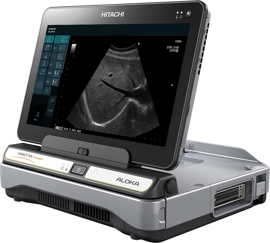
The new Hitachi Aloka Prologue is a touch screen, full color, doppler capable diagnostic ultrasound.
Abdominal
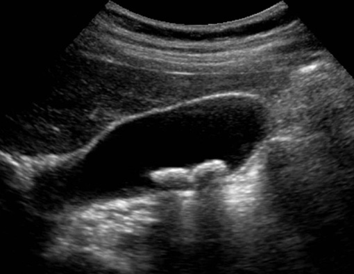
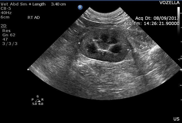
Below are images of the urinary bladder and kidney via ultrasound.

URINARY BLADDER WITH STONES
At Urban Vet we routinely use the Ultrasound for guided urine collection. This allows us to obtain a sterile sample as well as examine the bladder wall and visualize masses or stones within the bladder simultaneously. This is the safest, most effective way to collect urine in our pets.

Kidney - Transverse View
It is common to visualize the kidneys during an ultrasound. Occasionally we can see stones or masses within the organ. It is also a landmark by which to locate the very small adrenal glands. Adrenal glands can secrete excess hormone causing hair loss, weight gain and urinary abnormalities- commonly referred to as Cushing's Disease.
